Advancing care for Painful Diabetic Peripheral Neuropathy
The Neuropathy Recovery Initiative (NRI) is a pioneering translational research initiative developing and clinically evaluating a structured, evidence-informed treatment protocol for people living with painful diabetic peripheral neuropathy.
Our goal is to establish a new therapeutic pathway that addresses neuropathic pain and its wider effects on physical function, sleep, emotional well-being, and quality of life.

THE EVERYDAY BURDEN
Neuropathy can affect nearly every part of daily life
Diabetic peripheral neuropathy (DPN) is one of the most common long-term complications of diabetes and may affect up to half of people with diabetes over their lifetime [1,2]. Its impact can extend far beyond altered sensation [1–4]:
Pain and sensation: burning, stabbing or electric-shock pain, tingling, numbness, and sensitivity to touch.
Mobility and physical function: reduced balance, walking confidence, exercise tolerance, and ability to remain active.
Sleep and fatigue: difficulty sleeping, repeated waking, and reduced daytime energy.
Work and independence: challenges with physical tasks, concentration, productivity, and everyday activities.
Emotional well-being: anxiety, depression, frustration, fear of progression, and loss of confidence.

Evidence of the broader impact: A 2025 study of 6,960 people with diabetes found that DPN—particularly when accompanied by neuropathic pain—was associated with poorer quality of life and mental health [3].
WIDER CLINICAL CONSEQUENCES
When loss of sensation becomes a serious health risk
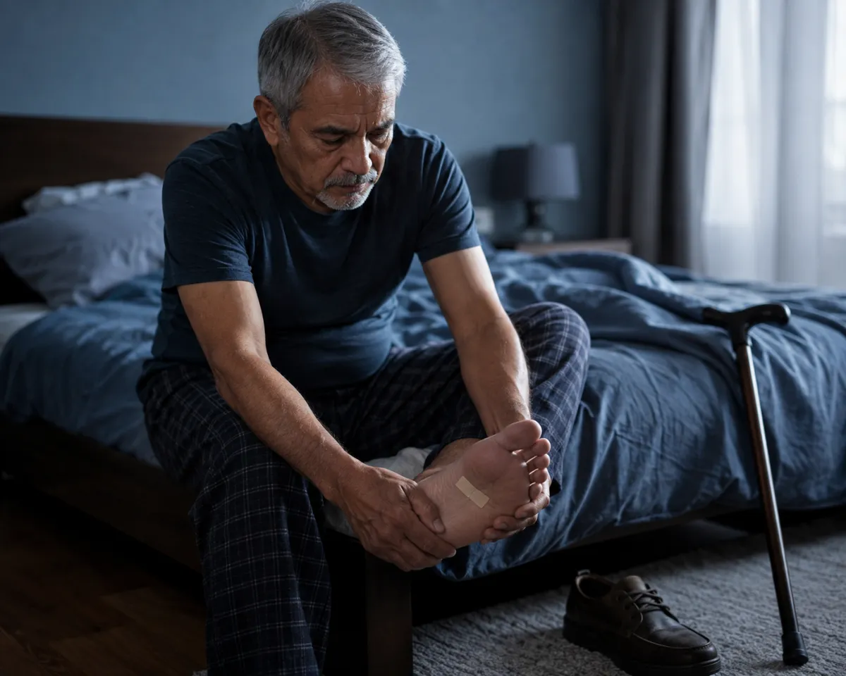
When DPN reduces protective sensation, cuts, blisters, and pressure injuries may go unnoticed. Combined with impaired circulation, immune function, and wound healing, these injuries can progress to foot ulceration, infection, hospitalization, and amputation [1,5].
Diabetic foot complications can have grave long-term consequences. A major clinical review reported five-year mortality rates of approximately 30% among people with diabetic foot ulcers and more than 70% among those who undergo major amputation [5].
These outcomes arise from multiple interacting conditions—not neuropathy alone—but illustrate how an initially unnoticed injury can contribute to a life-altering and potentially life-threatening chain of events.
THE UNMET NEED
Established treatments exist, but significant needs remain
Current care remains essential. Yet persistent symptoms and treatment limitations reinforce the need for research into more effective, integrated approaches that may extend beyond symptom control.
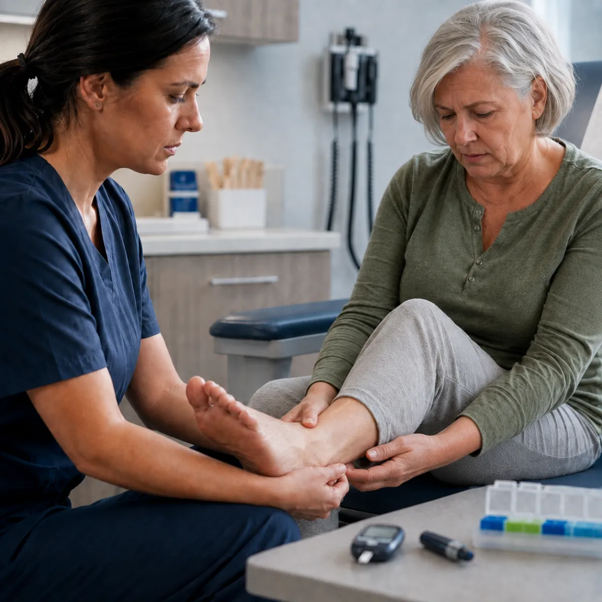
Management of diabetic peripheral neuropathy may include glucose and cardiovascular risk management, foot surveillance, physical activity, lifestyle support, and medications for neuropathic pain [6]. Several recommended medication classes can provide meaningful relief for some people [6].
However, no single treatment works for everyone. Pain relief is often partial, individual responses vary, and adverse effects—including dizziness, sedation, swelling, gastrointestinal symptoms, and cognitive effects—may limit treatment. Most available therapies primarily manage symptoms rather than repair or reverse established peripheral nerve damage [2,6,7].
A GROWING SYSTEMIC BURDEN
A global challenge with growing consequences
589 million
Adults worldwide living with diabetes in 2024 [50]
20-50%
Estimated DPN prevalence among people with type 2 diabetes [51]
20-40 %
Estimated painful DPN prevalence among people already affected by DPN [51]

Together, these figures point to a vast and expanding population at risk. By 2050, the number of adults living with diabetes worldwide is projected to reach 853 million, making the need for earlier detection, more effective symptom management, and improved care increasingly urgent [50]. The consequences extend across individuals, families, workplaces, and healthcare systems. In a survey of 506 adults receiving treatment for painful DPN, 66% reported disability related to nerve pain [52]. At the population level, DPN is associated with greater healthcare utilization and direct medical costs, while neuropathy-related disability contributes to lost productivity, reduced independence, and caregiving needs [51].
Taken together, the scale of DPN, its contribution to chronic disability and economic costs, and the persistence of unmet need make the search for more effective solutions an increasingly important priority for healthcare systems.
translational research for CLINICAL INNOVATION
Turning scientific findings
into Clinical Progress
Promising discoveries do not become better care automatically. Translating scientific findings into clinical progress is a multistage process that can be slowed by scientific, operational, regulatory, and organizational barriers [8,53]. Progress depends on purposeful collaboration across disciplines and sectors—connecting researchers, clinicians, patients, delivery partners, and health systems around a shared pathway from evidence to care [53].
Moving promising evidence forward requires more than recognizing its potential. It requires bringing evidence streams together, testing them rigorously, designing for real-world delivery, and integrating clinical expertise and patient priorities from the outset. Translational and implementation science provide the principles; impact comes from applying them to develop solutions that can be evaluated, delivered, adopted, and sustained in practice [53,54].
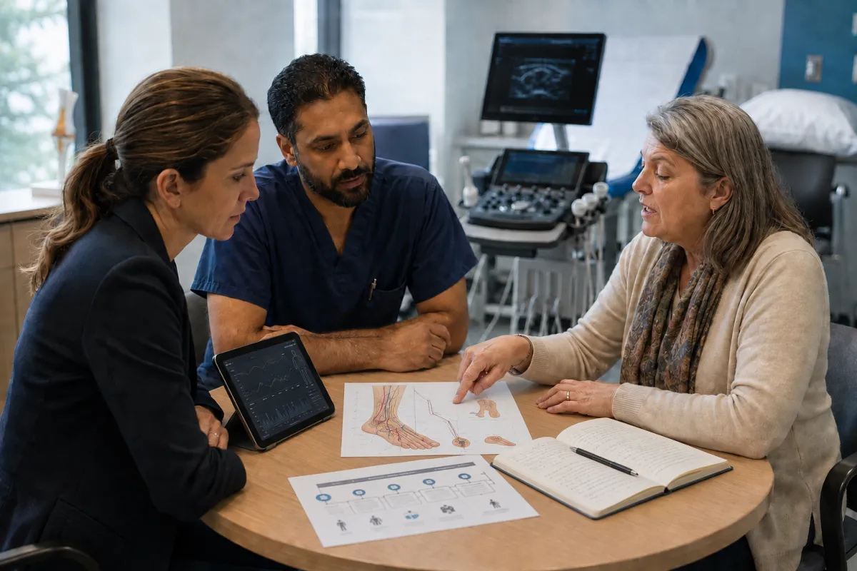
The Neuropathy Recovery Initiative is designed to bring scientific, clinical, patient, and delivery partners together to transform promising evidence into a testable model of care, embed clinical evaluation in real-world settings, and build a pathway toward a validated, scalable clinical service for painful DPN.
CONNECTING THE EVIDENCE
Four Evidence Streams.
One Integrated Clinical Innovation.
The Neuropathy Recovery Initiative is connecting four complementary evidence streams to build a standardized clinical protocol within a coherent, professionally delivered model of care designed around the complex needs of people with painful diabetic peripheral neuropathy.
EVIDENCE STREAM 01
Education & Self-Regulation
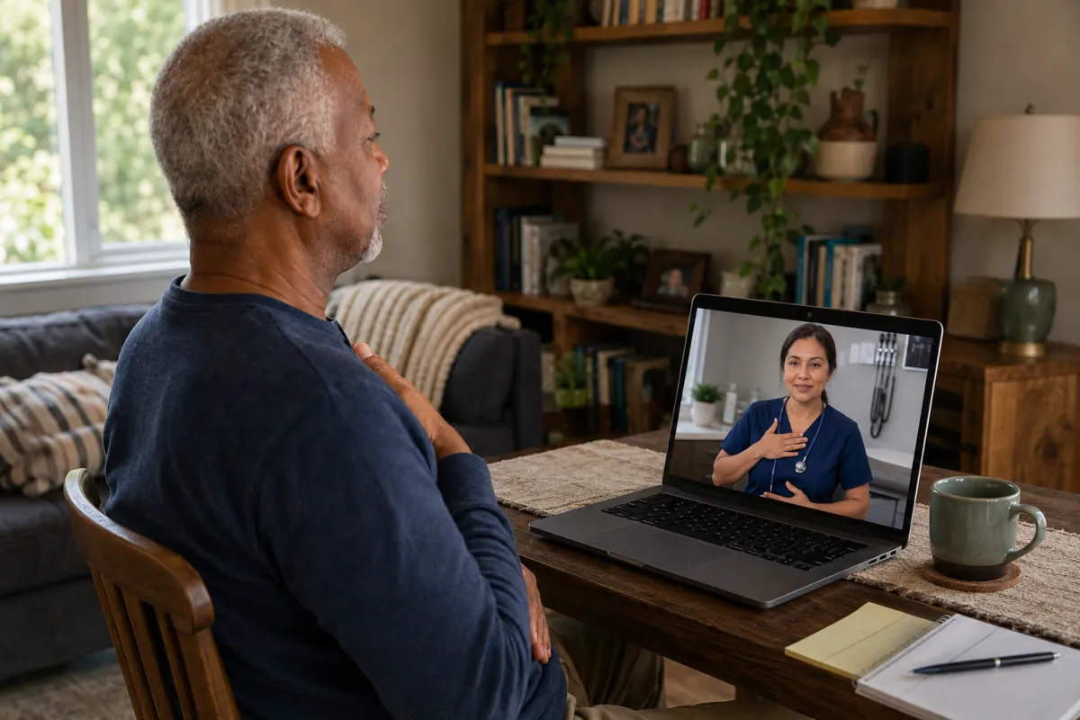
Painful diabetic neuropathy affects more than pain alone, often disrupting sleep, movement, emotional well-being, confidence, and participation in everyday life [3,4]. Evidence from educational, self-management, cognitive-behavioural, and mindfulness-based interventions suggests that structured support can improve pain-related outcomes, self-efficacy, quality of life, and management of neuropathy-related symptoms and complications [36–38,55].
NRI integrates education and guided self-regulation within a whole-person model of care, helping participants understand their condition, engage actively with treatment, strengthen practical self-management skills, and address the wider impact of painful DPN.
EVIDENCE STREAM 02
Bee Venom

Bee venom is a biologically complex candidate with a growing research foundation across neuropathic pain, pain modulation, neuroimmune activity, neural repair, vascular signalling, and tissue recovery [9–20,39,56–62]. Human evidence now extends beyond case observations, including controlled studies in chronic pain, preliminary randomized evidence in neuropathic pain, clinically measurable effects following topical application, and professional administration through injection and ultrasound-assisted delivery. Bee-venom phonophoresis has also been investigated in people with diabetic foot ulcers, with a randomized controlled trial reporting greater wound reduction than therapeutic ultrasound alone [46,56–62].
NRI brings these converging evidence streams into a standardized, professionally administered protocol to investigate whether bee venom can be translated safely and meaningfully to painful diabetic peripheral neuropathy—while addressing critical questions of formulation, dose, delivery, safety, reproducibility, and clinical impact.
EVIDENCE STREAM 03
Bee Propolis
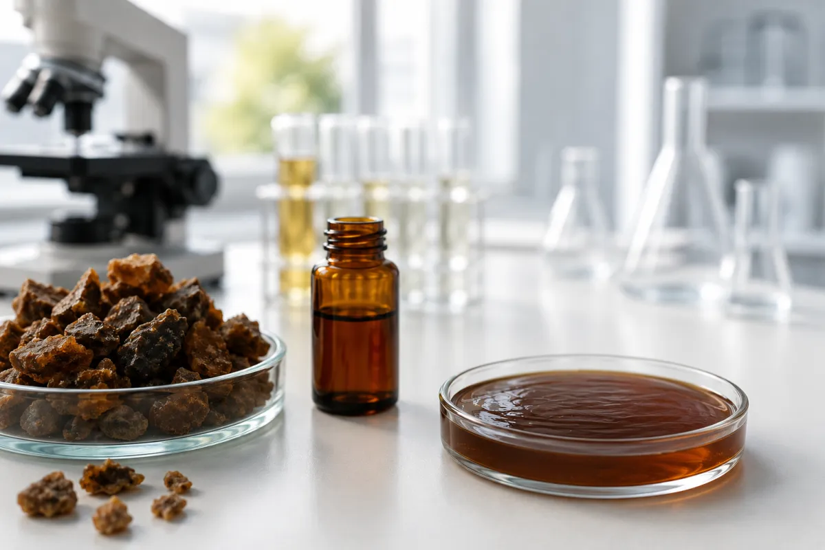
Propolis is a biologically diverse candidate with research spanning diabetes, inflammation, oxidative stress, experimental peripheral-nerve injury, vascular biology, antimicrobial activity, and tissue repair [21–26,41,42]. Human studies have also evaluated topical propolis directly in diabetic foot ulcers, reporting signals across wound reduction, tissue repair, inflammatory activity, and bacterial burden [43–45].
NRI investigates propolis as a standardized complementary component of the formulation, building on its distinct but overlapping biological profile with bee venom and existing research combining both substances [28]. The central question is whether propolis can add safe and meaningful value to an integrated intervention for painful diabetic peripheral neuropathy.
EVIDENCE STREAM 04
Phonophoresis Delivery
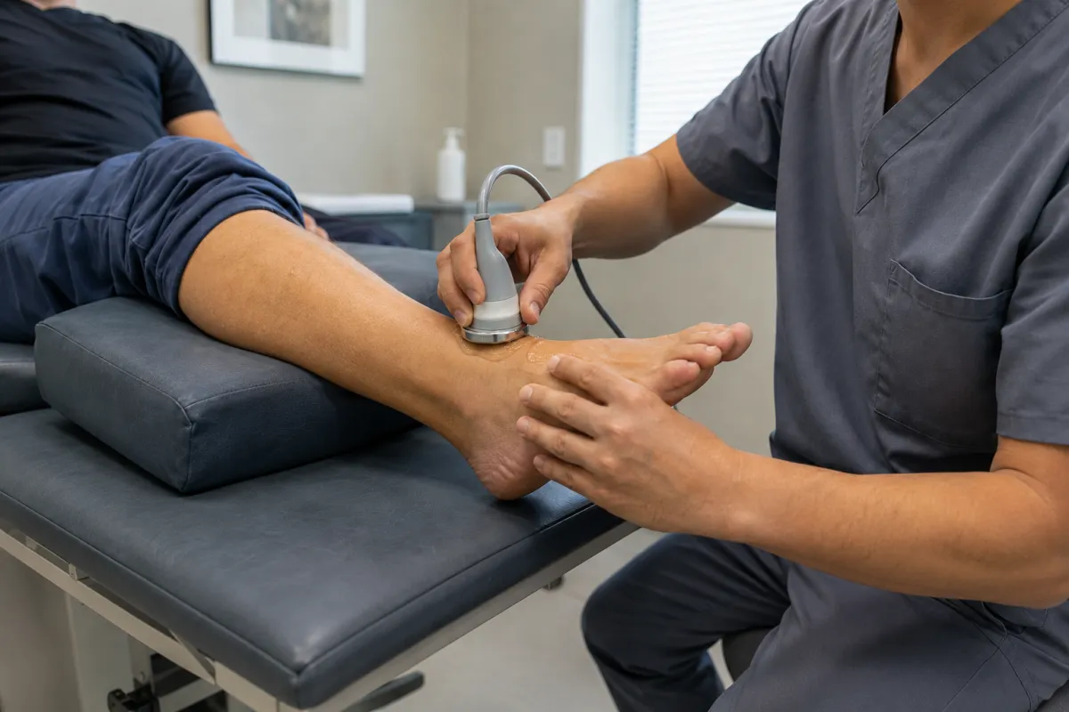
Phonophoresis uses therapeutic ultrasound to enhance the localized delivery of selected topical compounds, providing a clinically established platform in which treatment parameters and administration can be professionally controlled and standardized [29,30,48,49]. Human research has demonstrated enhanced local drug delivery for selected compounds, while bee-venom phonophoresis has produced clinical signals in controlled studies of postoperative recovery and diabetic and venous ulcers [46,58].
NRI investigates whether this delivery approach can translate a standardized bee-venom and propolis formulation into a safe, reproducible clinical procedure for painful DPN—while systematically evaluating formulation, ultrasound parameters, treatment frequency, safety, and clinical outcomes.
OUR TRANSLATIONAL PATHWAY
From Converging Evidence to Scalable Clinical Impact
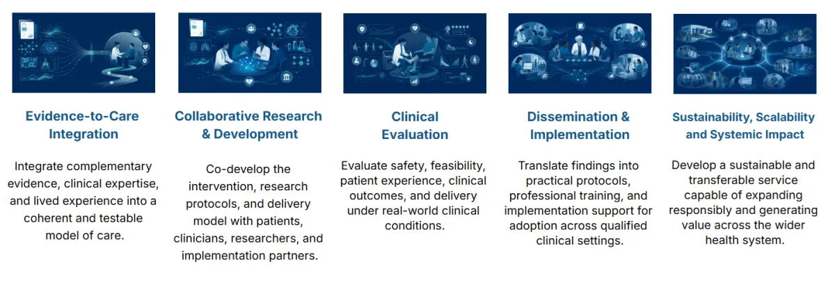
The Neuropathy Recovery Initiative brings together complementary scientific evidence, clinical expertise, lived experience, and implementation planning to develop an integrated model of care for painful diabetic peripheral neuropathy. Its translational pathway connects evidence integration, collaborative development, clinical evaluation, implementation, and responsible scale—supported by patient-centred leadership and multidisciplinary collaboration across research, clinical, technical, institutional, and community partners [53,54,66].
Each stage is shaped by the questions that determine real-world value: Can the model improve outcomes that matter to patients? Can trained professionals deliver it safely and consistently in clinical practice? And, if supported by the evidence, can it be standardized, implemented, and responsibly scaled as a sustainable clinical service?
"The objective is not research as an endpoint. It is to use research to build, validate, and prepare a new clinical service capable of generating sustainable impact for patients, professionals, healthcare organizations, and the wider health system."
Tiago Guardia, PhD - Initiative Founder
FROM EVIDENCE TO ACTION
Building Impact Through Collaboration
Our goal is to assemble the academic, clinical, community, and innovation capabilities required to develop a rigorous research program and translate its findings into meaningful clinical impact.
The Neuropathy Recovery Initiative has established a scientific rationale, an integrated clinical concept, and a clearly defined translational opportunity. The next stage is to build the academic and institutional foundation needed to develop and evaluate the model—while connecting scientific leadership with clinical expertise, lived experience, research infrastructure, technical development, and funding.

Academic Research Partners
NRI is seeking formal collaboration with academic researchers and institutions that can contribute scientific leadership, complementary expertise, research infrastructure, and the institutional foundation required for protocol development and multisite clinical investigation.
Clinicians and Healthcare Systems
Clinicians, healthcare organizations, and prospective research sites can contribute clinical oversight, participant pathways, operational expertise, and real-world care environments in which the proposed intervention can be evaluated and progressively refined.
People Living with Neuropathy and Community Partners
People with lived experience, caregivers, advocates, and community organizations can help ensure that the research reflects the everyday realities of neuropathy and prioritizes outcomes that are meaningful to the people it is intended to serve.
Industry, Innovation and Funding Partners
NRI has established an investigational formulation concept and an early prototype foundation. Selected partners can contribute specialized capabilities in formulation testing, quality assurance, manufacturing, regulatory strategy, technical development, and research funding—supporting the rigorous evaluation of the intervention and, if justified by the evidence, its progression toward clinical implementation.
A MESSAGE FROM THE INITIATIVE LEAD
Beyond Discovery: Making Impact Real
Healthcare innovation depends on more than scientific discovery and technological development. It requires vision and collective capacity to connect evidence with practice and technical capabilities with clinical needs, so that innovation can address systemic challenges and improve people’s lives.
Too often, promising findings remain separated across disciplines, institutions, and sectors, while translation and implementation are considered only after the research is complete. Closing this distance requires a more connected approach, in which research, care, implementation, data, and continuous improvement shape and strengthen one another from the beginning.
Through this initiative, we bring together scientific evidence, an integrated intervention, multisite clinical evaluation, patient education, professional training, and implementation planning within a single translational pathway. This work responds to the profound burden of painful diabetic peripheral neuropathy on individuals and families, clinical services, healthcare systems, workforce participation, and the wider economy.
Our objective is to conduct research designed from the outset to generate real-world impact and build an operational model for clinical service. If the model demonstrates sufficient safety, feasibility, and clinical value, the research can provide a direct foundation for continued service delivery at participating sites and replication across additional clinical settings.
In advancing a much-needed response to painful diabetic neuropathy, we can also demonstrate how cross-sector collaboration can move scientific potential more effectively into practice.
The distance between what science makes possible and what people experience in care will not close by itself. It closes when researchers, clinicians, communities, healthcare organizations, public institutions, innovators, and funders choose to build the pathway together.
If you see a role for your knowledge, experience, resources, or leadership in making this happen, I invite you to bring your contribution forward. Together, we can move beyond discovery, advance a new response to painful diabetic neuropathy, and build a model for turning scientific potential into real-world impact.

Tiago Guardia, PhD
Initiative Founder
STAY CONNECTED
Follow the Research.
Help Shape What Comes Next.
The Neuropathy Recovery Initiative is entering a partnership-building and research development phase. Whether you are interested in contributing to its development or following its progress, there is a way to take part.
Explore Collaboration
Interested in contributing expertise, sharing lived experience, exploring an academic, clinical, institutional, or industry partnership, or supporting the development of the research program? We welcome the conversation.
Follow the Initiative
Join our mailing list to receive occasional updates on research progress, new partnerships, publications, and opportunities to contribute or become involved.
** Joining the mailing list does not constitute enrolment in a research study. Please do not submit personal medical information through this form.
Scientific Evidence
DETAILED EVIDENCE-BASED INFORMATION ABOUT THE TOPICS
Whole-Person Approach
Education, self-management, and self-regulation beyond the procedure
Painful diabetic peripheral neuropathy affects considerably more than pain intensity. It can disrupt sleep, movement, emotional well-being, confidence, relationships, work, and participation in everyday life [3,4]. A treatment model focused exclusively on the procedure may therefore leave important dimensions of the patient experience unaddressed.
NRI proposes a whole-person framework in which structured education, pain self-management, and guided self-regulation complement the investigational procedure. The objective is not to position these approaches as substitutes for medical care, but to help participants better understand their condition, participate actively in treatment, and develop practical skills for managing its effects in everyday life.
Education and active self-management
Diabetes self-management education and support is already an established component of diabetes care, with demonstrated benefits for clinical outcomes, psychosocial well-being, and quality of life [31,36]. NRI would build on this foundation while focusing specifically on the challenges created by painful diabetic neuropathy.
The education component may include neuropathy awareness, foot protection, symptom monitoring, treatment communication, movement and activity pacing, sleep support, and preparation for the investigational procedure. It may also incorporate practical pain-management strategies informed by cognitive-behavioural and other evidence-based self-management approaches.
Importantly, clinical research has begun to evaluate education specifically in people with painful diabetic neuropathy. Educational interventions have reported improvements in pain management, self-efficacy, and quality of life, while broader evidence supports health education for self-care and management of neuropathy-related symptoms and complications [36,55].
This creates an opportunity for NRI to develop education not simply as supporting information, but as a structured and measurable component of the clinical model.
Pain, behaviour, and emotional well-being
Chronic neuropathic pain is experienced within a wider interaction among physical symptoms, behaviour, sleep, stress, mood, expectations, and everyday activity. Psychological approaches do not imply that neuropathic pain is psychological in origin; rather, they provide tools for reducing the additional burden created by persistent pain and improving the ways people respond to and live with symptoms.
A systematic review and meta-analysis of randomized trials in diabetic neuropathy found that cognitive-behavioural and mindfulness-based interventions were associated with improvements in pain severity and quality of life [37]. These findings support evaluating behavioural and psychological outcomes alongside biological and clinical outcomes within NRI.
Mind–body self-regulation
Mindfulness meditation and related mind–body practices may provide additional tools for responding to pain, stress, difficult emotions, and sleep disruption.
A 2024 systematic review and meta-analysis of meditation-based interventions for chronic peripheral neuropathy reported improvements in anxiety, depression, mindfulness, and pain severity at follow-up [32]. Broader randomized-trial evidence has also associated mindfulness meditation with modest improvements in chronic pain, depression, and quality of life [33].
Yoga Nidra, a guided meditation and deep-relaxation practice, provides another potentially accessible approach to self-regulation. Emerging evidence suggests potential benefits across pain, psychological well-being, stress regulation, and sleep. A systematic review and meta-analysis reported reductions in pain compared with passive control conditions, while recent randomized research has also examined effects on psychological well-being and physiological stress regulation [63,64].
Within NRI, these practices could be evaluated for their effects on pain coping, sleep, stress regulation, emotional well-being, self-efficacy, physical function, and quality of life.
From patient education to an integrated care experience
The whole-person component creates an opportunity to make participation in NRI more than a sequence of treatment sessions. Assessment, education, the investigational procedure, self-management support, and outcome monitoring can be integrated into a coherent clinical experience designed around the needs of people living with painful DPN.
This approach also creates measurable research questions. Alongside pain and clinical outcomes, NRI can examine changes in self-efficacy, treatment engagement, sleep, emotional well-being, physical function, self-management, and quality of life.
The translational opportunity is therefore not only to investigate a new procedure, but to determine whether that procedure can be embedded within a structured, human-centred model of care that helps people understand their condition, participate actively in treatment, and better manage its impact on everyday life.
Bee Venom
A multidimensional candidate for neuropathy research
Bee venom is a complex natural substance containing biologically active peptides and enzymes, including melittin, apamin, and phospholipase A2. Rather than acting through a single biological target, these components have been investigated across pain signalling, inflammatory and immune pathways, neural communication, vascular signalling, tissue repair, and antimicrobial activity [9,10].
This breadth of activity makes bee venom a compelling translational research candidate for painful diabetic peripheral neuropathy—a condition shaped by interacting sensory, inflammatory, metabolic, vascular, and neurodegenerative processes.
Neuropathic-pain modulation
In animal models of peripheral neuropathic pain, diluted bee-venom interventions have reduced mechanical allodynia, thermal hyperalgesia, and cold allodynia. Mechanistic studies have identified the involvement of adrenergic, noradrenergic, serotonergic, sensory-receptor, immune, and spinal excitatory-signalling pathways [11–14,62].
These findings are particularly relevant to painful DPN because they suggest that bee venom may influence several levels of pain processing—from peripheral sensory mechanisms to spinal signalling and descending pain modulation.
Human evidence across pain conditions
Human research extends beyond isolated case observations. Bee-venom interventions have been evaluated across a range of painful conditions, most commonly through injection of diluted bee venom at acupuncture points. An updated systematic review and meta-analysis identified 20 randomized controlled trials of bee-venom acupuncture for musculoskeletal pain. Pooled analyses favoured bee venom over sham saline injection in several comparisons, although small samples, variation in dose and concentration, and clinical heterogeneity limit definitive conclusions [59].
Individual controlled trials provide additional evidence. In chronic low-back pain, a randomized double-blind sham-controlled trial found greater improvements in pain-related bothersomeness, pain intensity, and functional status following bee-venom acupuncture than sham treatment [60]. Clinical experience across other musculoskeletal and inflammatory pain conditions further supports continued investigation of bee venom as a pain-modulating intervention [20,39,59,62].
Importantly, preliminary human evidence also extends to neuropathic pain. In a small randomized controlled trial involving central post-stroke pain—a neuropathic pain syndrome—acupoint injections of diluted bee venom produced greater pain reduction than saline injections after three weeks of treatment [57]. Case reports have additionally described improvement following bee-venom-based treatment in refractory postherpetic neuralgia and in a patient with diabetic neuropathy receiving combined Korean medicine [15,16].
These studies do not establish efficacy in painful DPN. They do, however, provide a human clinical bridge between experimental evidence of bee-venom-mediated pain modulation and the specific neuropathic-pain hypothesis proposed for NRI.
Topical delivery and clinical activity
Bee venom has also demonstrated clinically measurable effects when administered topically without injection or ultrasound.
In a randomized double-blind study, a topical ointment containing bee venom was applied over the masseter muscles of patients with temporomandibular myofascial pain. Treatment was associated with reductions in muscle tension, providing controlled human evidence that a topical bee-venom formulation can produce biological effects without injection [56].
A larger randomized, double-blind, multicentre study involving 136 patients with atopic dermatitis compared a bee-venom-containing emollient with an otherwise similar vehicle. The bee-venom group showed greater improvements in disease severity and pruritus [61].
These studies involve conditions very different from DPN and do not demonstrate delivery to peripheral nerves. Nevertheless, they provide important evidence that bee-venom-containing formulations can exert clinically detectable effects following passive topical administration in humans.
Small nerve fibres and neural repair
One of the most intriguing findings comes from a mouse model of chemotherapy-induced peripheral neuropathy. Repeated diluted bee-venom treatment reduced mechanical allodynia and was associated with restoration of intraepidermal nerve-fibre loss [13].
These small sensory fibres transmit pain and temperature information and are frequently affected early in DPN. Their restoration in an experimental neuropathy model raises an important question for future research: could bee-venom-based interventions influence nerve integrity as well as pain?
Supporting this possibility, apamin—a peptide present in bee venom—has promoted neurite outgrowth and axonal regeneration following experimental neuronal injury, accompanied by increased expression of brain-derived neurotrophic factor, nerve growth factor, and regeneration-associated genes [17].
Inflammatory and neuroimmune pathways
Persistent inflammation and altered neuroimmune signalling contribute to diabetic nerve injury and neuropathic pain [2]. Experimental research has found that bee venom and its components can influence inflammatory cytokines, cyclooxygenase pathways, NF-κB-related signalling, microglial activity, macrophage function, and regulatory immune responses [9,10,62].
This does not establish a clinical anti-inflammatory effect in DPN, but it provides a mechanistic foundation for evaluating inflammatory and neuroimmune biomarkers alongside pain and functional outcomes.
Vascular, oxidative, and tissue-repair signalling
Diabetic neuropathy does not occur in isolation from the wider metabolic and vascular environment. Impaired microvascular function, oxidative stress, and reduced tissue resilience can contribute to nerve dysfunction and increase the consequences of unnoticed injury.
In diabetic mouse wound models, bee venom accelerated wound closure and influenced multiple processes involved in tissue repair. Reported findings included increased collagen production; restoration of VEGF and TGF-β signalling; endothelial-progenitor-cell recruitment; increased CD31-positive neovascularization; improved macrophage survival; and modulation of Ang-1/Tie-2, eNOS, AKT, ERK, and Nrf2 pathways [18,19].
Bee venom and melittin have also demonstrated antibacterial, antifungal, and antibiofilm activity in laboratory research [9,10,47]. In diabetic-wound models, bee venom increased β-defensin-2 expression and supported cellular responses relevant to wound defence [19].
Wound healing and neuropathy remain distinct clinical targets, and laboratory antimicrobial activity does not establish treatment of diabetic-foot infection. However, these findings broaden the biological rationale for investigating vascular, inflammatory, oxidative, tissue-repair, and tissue-defence pathways within the wider diabetic-foot research landscape.
Professional phonophoresis and human diabetic-wound research
Therapeutic ultrasound provides another clinically investigated route for bee-venom administration.
In a randomized controlled trial following inguinal hernia surgery, 66 participants received low-intensity pulsed ultrasound with either bee-venom gel or plain gel. After three weeks, the bee-venom phonophoresis group showed greater improvements in pain, C-reactive protein, and hip range of motion [58]. Although this was an acute postoperative rather than neuropathic-pain population, the study provides controlled human evidence for professionally administered bee venom through ultrasound-assisted topical delivery.
More directly relevant to the diabetic-foot setting, bee-venom phonophoresis has been evaluated in people with chronic diabetic foot ulcers and venous ulcers. A single-blind randomized controlled trial included 100 participants assigned across four groups. Participants received standard wound care combined with either therapeutic ultrasound alone or phonophoresis using a bee-venom gel. After 12 weeks, the bee-venom phonophoresis groups showed greater reductions in wound surface area and ulcer volume than their respective control groups. Tissue analysis also identified increased Ki-67 expression, indicating greater cellular proliferation within granulation tissue [46].
This study provides important early clinical evidence that bee venom can be administered through phonophoresis within professional diabetic wound-care settings. Together with broader research on topical bee venom and professionally administered bee-venom interventions, it provides a foundation for investigating formulation standardization, ultrasound parameters, treatment frequency, safety, biological mechanisms, and reproducibility.
Because the diabetic-wound evidence currently rests on a limited clinical literature, independent confirmation is needed. Nevertheless, the relationship among bee venom, ultrasound-assisted delivery, and diabetic tissue pathology has progressed beyond experimental rationale to human clinical investigation.
From biological promise to a standardized procedure
Taken together, the evidence presents bee venom not as a single-mechanism analgesic, but as a biologically complex candidate supported by converging research across pain modulation, small-fibre integrity, neuroimmune activity, vascular signalling, oxidative regulation, and tissue repair.
Human research adds another important dimension. Bee venom has been investigated through several delivery approaches—including acupoint injection, passive topical formulations, and ultrasound-assisted topical delivery—with controlled trials reporting clinical signals across neuropathic, musculoskeletal, inflammatory, dermatological, postoperative, and wound-related conditions [20,39,56–62].
None of this establishes that bee venom is safe or effective for painful diabetic peripheral neuropathy. Instead, it defines the translational opportunity: a biologically plausible intervention with experimental neuropathy evidence, an expanding human pain literature, demonstrated topical activity, and early clinical experience with ultrasound-assisted delivery, but without definitive controlled evaluation in painful DPN.
The central research questions therefore concern how bee venom should be characterized, standardized, formulated, dosed, delivered, and monitored. Previous clinical research has documented local and systemic adverse reactions, including the possibility of serious hypersensitivity, making ingredient characterization, allergy precautions, professional administration, and systematic adverse-event monitoring essential [20,40].
Taken together, this convergence of mechanistic evidence and early human clinical experience creates a clear research opportunity: to determine whether a standardized, professionally administered bee-venom intervention can translate into safe and meaningful benefits for people living with painful diabetic peripheral neuropathy.
Bee Propolis
A biologically diverse candidate for neuropathy research
Propolis is a resinous material produced by bees from botanical sources. It contains phenolic acids, flavonoids, terpenes, and other biologically active compounds whose composition varies with geography, season, and plant origin.
This natural diversity contributes to the biological potential of propolis, but it also makes chemical characterization and standardization essential for reproducible research. Standardized propolis preparations have been investigated across diabetes, inflammation, oxidative stress, antimicrobial activity, tissue repair, vascular biology, and experimental peripheral nerve injury [21–26,41,42].
Antioxidant and anti-inflammatory activity
Oxidative stress and persistent inflammation are closely connected to the metabolic, vascular, and neural processes involved in diabetic neuropathy.
In an 18-week randomized trial involving people with type 2 diabetes, Brazilian green propolis increased glutathione and total polyphenol levels while reducing protein carbonyls and lactate-dehydrogenase activity, indicating improved systemic antioxidant status [21].
A meta-analysis of 27 randomized trials similarly found that propolis supplementation reduced several inflammatory biomarkers, including C-reactive protein, interleukin-6, tumour necrosis factor-α, and MCP-1, while increasing measures of antioxidant capacity [22]. The magnitude and consistency of these effects varied across biomarkers, populations, doses, and propolis preparations.
These human findings do not establish an effect on diabetic neuropathy or demonstrate that the same biological effects would occur with topical administration. They do, however, show that standardized propolis preparations can influence inflammatory and oxidative pathways relevant to the diabetic environment in which peripheral nerve injury develops [21–23].
Experimental neuropathy and nerve repair
Direct experimental evidence has begun to connect propolis with diabetic peripheral neuropathy. In a streptozotocin-induced rat model, propolis—evaluated alone and in combination with quercetin—produced antioxidant, biochemical, histological, and neuroprotective effects in sciatic-nerve tissue [24].
Propolis has also been investigated in models of traumatic peripheral-nerve injury. A propolis–gum Arabic nerve-guidance channel improved functional recovery following sciatic-nerve transection and was associated with increased numbers of myelinated axons, larger myelinated fibres, and improved muscle-fibre outcomes [25]. A separate study found that hydroalcoholic red-propolis extract supported functional recovery and axonal repair following experimental sciatic-nerve injury [41].
Although traumatic nerve injury differs substantially from DPN, these findings broaden the research rationale beyond systemic metabolic effects and raise important questions about whether propolis-related antioxidant, anti-inflammatory, and tissue-protective activity could help protect peripheral nerves or support regenerative processes.
Vascular, tissue-repair, and antimicrobial activity
Microvascular dysfunction, oxidative injury, persistent inflammation, and impaired tissue repair contribute to diabetic nerve damage and to the wider consequences of diabetic foot disease.
In cultured human endothelial cells, Brazilian green propolis reduced oxidized-LDL-induced oxidative injury through signalling involving PI3K/Akt/mTOR and Nrf2/HO-1 [26]. Propolis-containing experimental dressings and biomaterials have also demonstrated antioxidant, antimicrobial, collagen-supporting, angiogenic, and re-epithelialization effects across laboratory and animal wound models [42].
Propolis preparations have additionally demonstrated antibacterial, antifungal, and antibiofilm activity in laboratory studies. Human research in oral health has reported reductions in plaque accumulation and gingival inflammation with some propolis formulations [27], providing evidence that propolis can retain biological activity within topical and mucosal preparations.
These findings do not establish a role for propolis in preventing or treating diabetic-foot infection. Any such indication would require formulation-specific antimicrobial testing and dedicated clinical evaluation. They do, however, strengthen the rationale for investigating tissue resilience, vascular function, and wound-related outcomes within the broader diabetic-foot research landscape.
Human diabetic-foot-ulcer research
Importantly, topical propolis has progressed beyond experimental wound models and has been evaluated directly in people with diabetic foot ulcers.
In a prospective feasibility study, 24 participants with longstanding diabetic foot ulcers received topical propolis alongside standard wound care. Compared with a matched control cohort, propolis-treated ulcers showed greater reductions in wound area, lower bacterial counts, reduced activity of the inflammatory enzyme MMP-9, and higher rates of complete healing at selected follow-up points. No propolis-related adverse effects were reported [43].
A randomized controlled trial involving people with Wagner grade 1 or 2 diabetic foot ulcers found that adding topical propolis ointment to conventional treatment produced a greater reduction in ulcer size over four weeks than conventional treatment alone. Differences in several secondary inflammatory and wound measures were not statistically significant [44].
A separate clinical study evaluating propolis spray as an adjunct to standard diabetic-foot-wound care reported greater wound-area reduction and increased connective-tissue deposition in the propolis group [45].
Together, these studies provide early human evidence for topically administered propolis within the diabetic-foot setting [43–45]. Larger and more rigorous multicentre trials are needed to establish efficacy, identify appropriate standardized preparations and doses, and determine which wounds and patient populations are most likely to benefit.
Scientific rationale for combination with bee venom
Propolis is particularly interesting to NRI not only as an independent bioactive candidate, but as a potentially complementary component of the investigational formulation.
Bee venom and propolis have distinct but partially overlapping research profiles across inflammatory regulation, oxidative stress, antimicrobial activity, vascular biology, and tissue repair. Published research has also evaluated bee venom and propolis together within the same topical formulation, providing precedent for investigating their combined use [28].
This does not establish synergy between the two substances or demonstrate that their combination will improve neuropathic outcomes. Determining whether propolis contributes additional biological or clinical value to a standardized bee-venom formulation is itself an important translational research question.
From biological activity to a standardized formulation
Taken together, the evidence identifies propolis as a multidimensional research candidate supported by human metabolic and inflammatory research, experimental peripheral-nerve studies, vascular and tissue-repair findings, antimicrobial research, and early clinical investigation in diabetic foot ulcers.
Its natural chemical variability also creates an important development challenge. Botanical origin, extraction method, chemical composition, concentration, formulation, and stability must be characterized if propolis is to become part of a reproducible clinical intervention.
For NRI, the research opportunity is therefore to determine whether a chemically characterized and standardized propolis preparation can provide complementary value within a professionally delivered intervention for painful diabetic peripheral neuropathy.
Therapeutic Ultrasound Delivery
A controlled platform for localized treatment
Phonophoresis uses therapeutic ultrasound to enhance the movement of selected topical compounds into or through the skin. Ultrasound-mediated delivery has been investigated for decades, with mechanisms and delivery efficiency influenced by ultrasound frequency, intensity, duty cycle, treatment duration, application technique, formulation properties, treatment area, skin characteristics, and the molecular properties of the compounds being delivered [29].
This creates an important translational advantage for NRI. Rather than relying on variable self-application of a topical formulation, therapeutic ultrasound provides a professionally administered platform in which treatment parameters, formulation exposure, treatment area, frequency of administration, and clinical monitoring can be standardized and systematically evaluated.
Evidence for enhanced topical delivery
Human research demonstrates that ultrasound can alter the local delivery of selected topical compounds.
In a human pharmacokinetic study, phonophoresis with ketoprofen produced higher local tissue concentrations than passive topical application while plasma concentrations remained low [48]. Clinical studies using pharmaceutical gels have also reported benefits with phonophoresis across musculoskeletal conditions [30,49].
A systematic review and meta-analysis of nine randomized controlled trials involving 423 participants with knee osteoarthritis found improvements in pain or function with several phonophoresis interventions compared with therapeutic ultrasound using placebo gel, although results varied according to the drug and outcome evaluated [30].
These findings do not demonstrate that ultrasound will enhance delivery of bee-venom peptides or propolis constituents to the clinically relevant targets in painful DPN. Delivery is formulation- and molecule-dependent. They do, however, establish phonophoresis as a clinically investigated approach to localized topical drug delivery and provide a foundation for formulation-specific research within NRI.
Ultrasound and painful diabetic neuropathy
Therapeutic ultrasound may also have relevance beyond its role as a delivery technology.
A 2025 randomized placebo-controlled pilot study involving 50 people with painful diabetic peripheral neuropathy evaluated low-intensity focused ultrasound as a treatment for neuropathic pain. Participants receiving active ultrasound showed greater improvements in pain and neuropathic symptom measures than those receiving placebo treatment, although nerve-conduction measures did not significantly improve and larger, longer studies are needed [65].
Low-intensity focused ultrasound is technically and mechanistically distinct from the therapeutic ultrasound parameters proposed for phonophoresis, so these findings cannot be directly transferred to the NRI protocol. Nevertheless, the study provides early human evidence that ultrasound-based interventions are being investigated directly within the painful-DPN population and raises an additional research question regarding the contribution of ultrasound itself to clinical outcomes.
Professional phonophoresis with bee venom
The delivery concept proposed by NRI also has direct human clinical precedent with bee venom.
In a randomized controlled trial following inguinal hernia surgery, low-intensity pulsed ultrasound combined with bee-venom gel produced greater improvements in pain, C-reactive protein, and hip range of motion than ultrasound with plain gel [58].
Bee-venom phonophoresis has also been evaluated in people with chronic diabetic foot ulcers and venous ulcers. In a randomized controlled trial involving 100 participants, standard wound care combined with bee-venom phonophoresis produced greater reductions in wound surface area and ulcer volume than standard care combined with therapeutic ultrasound alone. Histological analysis also demonstrated increased Ki-67 expression within granulation tissue [46].
These studies involve clinical conditions different from painful DPN and do not establish efficacy for neuropathic pain. However, they demonstrate that professionally administered bee-venom phonophoresis has progressed beyond theoretical drug-delivery rationale to controlled human clinical investigation—including within the diabetic-foot setting.
Professional administration, standardization, and safety
Professional delivery is particularly important when the investigational formulation contains bee venom.
Clinical research with bee-venom interventions has documented local reactions including redness, swelling, itching, and discomfort, while systemic allergic reactions are less common but can be serious [20,40]. Structured participant screening, standardized formulation and dosing, clinical observation, predefined stopping criteria, and appropriate emergency procedures are therefore important elements of protocol development.
Professional administration also creates opportunities to standardize treatment fidelity across research sites. Ultrasound parameters, treatment area, application technique, formulation quantity, session duration, treatment frequency, adverse-event monitoring, and participant response can all be defined prospectively and documented across sessions.
These characteristics make phonophoresis relevant not only as a potential delivery enhancer, but as a platform for developing a reproducible clinical procedure.
From delivery technology to a standardized clinical procedure
The central NRI hypothesis is not that therapeutic ultrasound automatically makes a topical formulation more effective. The critical questions are whether ultrasound meaningfully alters the delivery of the relevant bee-venom and propolis constituents, whether those changes translate into clinically meaningful outcomes, and whether the additional procedure provides sufficient value to justify its use.
Existing research provides several pieces of this translational rationale: established mechanisms of ultrasound-mediated transdermal delivery [29], human evidence of enhanced local delivery for selected pharmaceutical compounds [48], controlled clinical evidence across phonophoresis applications [30,49], preliminary ultrasound research directly in painful DPN [65], and human trials of bee-venom phonophoresis—including within diabetic wound care [46,58].
For NRI, the research opportunity is to bring these evidence streams together and determine whether therapeutic ultrasound can provide a safe, controlled, reproducible, and clinically meaningful delivery platform for a standardized bee-venom and propolis intervention in painful diabetic peripheral neuropathy.
List of Scientific References
1. Hicks, C. W., & Selvin, E. (2019). Epidemiology of peripheral neuropathy and lower extremity disease in diabetes. Current Diabetes Reports, 19, 86. https://doi.org/10.1007/s11892-019-1212-8
2. Dubský, M., Sojáková, D., Fejfarová, V., & Jude, E. B. (2026). Diabetic peripheral neuropathy: New diagnostics and treatment perspectives. Drugs & Aging, 43, 29–48. https://doi.org/10.1007/s40266-025-01267-5
3. Borbjerg, M. K., Wegeberg, A.-M., Nikontovic, A., Mørch, C. D., Arendt-Nielsen, L., Ejskjaer, N., Brock, C., Vestergaard, P., & Røikjer, J. (2025). Understanding the impact of diabetic peripheral neuropathy and neuropathic pain on quality of life and mental health in 6,960 people with diabetes. Diabetes Care, 48(4), 588–595. https://doi.org/10.2337/dc24-2287
4. Alleman, C. J. M., Westerhout, K. Y., Hensen, M., Chambers, C., Stoker, M., Long, S., & van Nooten, F. E. (2015). Humanistic and economic burden of painful diabetic peripheral neuropathy in Europe: A review of the literature. Diabetes Research and Clinical Practice, 109(2), 215–225. https://doi.org/10.1016/j.diabres.2015.04.031
5. Armstrong, D. G., Tan, T.-W., Boulton, A. J. M., & Bus, S. A. (2023). Diabetic foot ulcers: A review. JAMA, 330(1), 62–75. https://doi.org/10.1001/jama.2023.10578
6. Price, R., Smith, D., Franklin, G., et al. (2022). Oral and topical treatment of painful diabetic polyneuropathy: Practice guideline update summary. Neurology, 98(1), 31–43. https://doi.org/10.1212/WNL.0000000000013038
7. Soliman, N., Moisset, X., Ferraro, M. C., et al. (2025). Pharmacotherapy and non-invasive neuromodulation for neuropathic pain: A systematic review and meta-analysis. The Lancet Neurology, 24(5), 413–428. https://doi.org/10.1016/S1474-4422(25)00068-7
8. Morris, Z. S., Wooding, S., & Grant, J. (2011). The answer is 17 years, what is the question: Understanding time lags in translational research. Journal of the Royal Society of Medicine, 104(12), 510–520. https://doi.org/10.1258/jrsm.2011.110180
9. Wehbe, R., Frangieh, J., Rima, M., El Obeid, D., Sabatier, J.-M., & Fajloun, Z. (2019). Bee venom: Overview of main compounds and bioactivities for therapeutic interests. Molecules, 24(16), 2997. https://doi.org/10.3390/molecules24162997
10. Zhang, S., Liu, Y., Ye, Y., Wang, X.-R., Lin, L.-T., Xiao, L.-Y., Zhou, P., Shi, G.-X., & Liu, C.-Z. (2018). Bee venom therapy: Potential mechanisms and therapeutic applications. Toxicon, 148, 64–73. https://doi.org/10.1016/j.toxicon.2018.04.012
11. Roh, D.-H., Kwon, Y.-B., Kim, H.-W., et al. (2004). Acupoint stimulation with diluted bee venom alleviates thermal hyperalgesia in a rodent neuropathic pain model: Involvement of spinal alpha-2-adrenoceptors. The Journal of Pain, 5(6), 297–303. https://doi.org/10.1016/j.jpain.2004.05.003
12. Kang, S.-Y., Roh, D.-H., Yoon, S.-Y., Moon, J.-Y., Kim, H.-W., Lee, H.-J., Beitz, A. J., & Lee, J.-H. (2012). Repetitive treatment with diluted bee venom reduces neuropathic pain via potentiation of locus coeruleus noradrenergic neuronal activity and modulation of spinal NR1 phosphorylation in rats. The Journal of Pain, 13(2), 155–166. https://doi.org/10.1016/j.jpain.2011.10.012
13. Yeo, J. H., Yoon, S. Y., Kwon, S. K., Kim, S. J., Lee, J. H., Beitz, A. J., & Roh, D. H. (2016). Repetitive acupuncture point treatment with diluted bee venom relieves mechanical allodynia and restores intraepidermal nerve fiber loss in oxaliplatin-induced neuropathic mice. The Journal of Pain, 17(3), 298–309.
https://doi.org/10.1016/j.jpain.2015.10.018
14. Lee, J.-H., Li, D. X., Yoon, H., Go, D., Quan, F. S., Min, B.-I., & Kim, S. K. (2014). Serotonergic mechanism of the relieving effect of bee venom acupuncture on oxaliplatin-induced neuropathic cold allodynia in rats. BMC Complementary and Alternative Medicine, 14, 471. https://doi.org/10.1186/1472-6882-14-471
15. Lee, S. M., Lim, J., Lee, J.-D., Choi, D.-Y., & Lee, S. (2014). Bee venom treatment for refractory postherpetic neuralgia: A case report. Journal of Alternative and Complementary Medicine, 20(3), 212–214.
https://doi.org/10.1089/acm.2013.0130 16. Jeon, G.-R., Yim, T.-B., Jeong, J.-Y., et al. (2021). A case report on diabetic neuropathy treated with bee venom acupuncture and combined Korean medicine. Journal of Internal Korean Medicine, 42(5), 958–966. https://koreascience.kr/article/JAKO202128837787061.page
17. Kim, H., Hong, J. Y., Lee, J., Jeon, W.-J., & Ha, I.-H. (2021). Apamin enhances neurite outgrowth and regeneration after laceration injury in cortical neurons. Toxins, 13(9), 603. https://doi.org/10.3390/toxins13090603
18. Badr, G., Hozzein, W. N., Badr, B. M., Al Ghamdi, A., Saad Eldien, H. M., & Garraud, O. (2016). Bee venom accelerates wound healing in diabetic mice by suppressing ATF-3 and iNOS-mediated oxidative stress and recruiting bone marrow-derived endothelial progenitor cells. Journal of Cellular Physiology, 231(10), 2159–2171. https://doi.org/10.1002/jcp.25328
19. Hozzein, W. N., Badr, G., Badr, B. M., Allam, A., Al Ghamdi, A., Al-Wadaan, M. A., & Al-Waili, N. S. (2018). Bee venom improves diabetic wound healing by protecting functional macrophages from apoptosis and enhancing Nrf2, Ang-1 and Tie-2 signaling. Molecular Immunology, 103, 322–335. https://doi.org/10.1016/j.molimm.2018.10.016
20. Jang, S., & Kim, K. H. (2020). Clinical effectiveness and adverse events of bee venom therapy: A systematic review of randomized controlled trials. Toxins, 12(9), 558. https://doi.org/10.3390/toxins12090558
21. Zhao, L., Pu, L., Wei, J., et al. (2016). Brazilian green propolis improves antioxidant function in patients with type 2 diabetes mellitus. International Journal of Environmental Research and Public Health, 13(5), 498. https://doi.org/10.3390/ijerph13050498
22. Bahari, H., Shahraki Jazinaki, M., Aliakbarian, M., Rashidmayvan, M., Golafrouz, H., Rahnama, I., Khodashahi, R., & Malekahmadi, M. (2025). Propolis supplementation on inflammatory and oxidative stress biomarkers in adults: A systematic review and meta-analysis of randomized controlled trials. Frontiers in Nutrition, 12, 1542184. https://doi.org/10.3389/fnut.2025.1542184
23. Zakerkish, M., Jenabi, M., Zaeemzadeh, N., Hemmati, A. A., & Neisi, N. (2019). The effect of Iranian propolis on glucose metabolism, lipid profile, insulin resistance, renal function and inflammatory biomarkers in patients with type 2 diabetes mellitus: A randomized double-blind clinical trial. Scientific Reports, 9, 7289. https://doi.org/10.1038/s41598-019-43838-8
24. Türedi, S., Çelik, H., Dağlı, Ş. N., Taşkın, S., Şeker, U., & Deniz, M. (2024). An examination of the effects of propolis and quercetin in a rat model of streptozotocin-induced diabetic peripheral neuropathy. Current Issues in Molecular Biology, 46(3), 1955–1974. https://doi.org/10.3390/cimb46030128
25. Nosratiyan, M., Farjah, G. H., Karimipour, M., & Pourheidar, B. (2021). The effect of propolis–gum Arabic as a novel nerve guidance channel on regeneration of sciatic nerve in male rats. Turkish Neurosurgery, 31(3), 361–367. https://doi.org/10.5137/1019-5149.JTN.29813-20.2
26. Yuan, W., Chang, H., Liu, X., et al. (2019). Brazilian green propolis inhibits Ox-LDL-stimulated oxidative stress in human umbilical vein endothelial cells partly through PI3K/Akt/mTOR-mediated Nrf2/HO-1 pathway. Evidence-Based Complementary and Alternative Medicine, 2019, 5789574. https://doi.org/10.1155/2019/5789574
27. Halboub, E., Al-Maweri, S. A., Al-Wesabi, M., Al-Kamel, A., Shamala, A., Al-Sharani, A., & Koppolu, P. (2020). Efficacy of propolis-based mouthwashes on dental plaque and gingival inflammation: A systematic review. BMC Oral Health, 20, 198. https://doi.org/10.1186/s12903-020-01185-5
28. Tanuğur Samancı, A. E., & Kekeçoğlu, M. (2022). Development of a cream formulation containing bee venom and other bee products. Journal of Cosmetic Dermatology, 21(10), 4913–4920. https://doi.org/10.1111/jocd.14891
29. Polat, B. E., Hart, D., Langer, R., & Blankschtein, D. (2011). Ultrasound-mediated transdermal drug delivery: Mechanisms, scope, and emerging trends. Journal of Controlled Release, 152(3), 330–348. https://doi.org/10.1016/j.jconrel.2011.01.006
30. Yang, F.-A., Chen, H.-L., Peng, C.-W., Liou, T.-H., Escorpizo, R., & Chen, H.-C. (2022). A systematic review and meta-analysis of the effect of phonophoresis on patients with knee osteoarthritis. Scientific Reports, 12, 12877. https://doi.org/10.1038/s41598-022-16084-8
31. Cochran, J., & Conn, V. S. (2008). Meta-analysis of quality of life outcomes following diabetes self-management training. The Diabetes Educator, 34(5), 815–823. https://doi.org/10.1177/0145721708323640
32. Babos, C. I., Leucuta, D. C., & Dumitrascu, D. L. (2024). Meditation-based therapies for chronic neuropathy: A systematic review and meta-analysis. Cureus, 16(8), e68226. https://doi.org/10.7759/cureus.68226
33. Hilton, L., Hempel, S., Ewing, B. A., et al. (2017). Mindfulness meditation for chronic pain: Systematic review and meta-analysis. Annals of Behavioral Medicine, 51(2), 199–213. https://doi.org/10.1007/s12160-016-9844-2
34. Datta, K., Tripathi, M., Verma, M., Masiwal, D., Mallick, H. N., & Kumar, V. M. (2021). Yoga nidra practice shows improvement in sleep in patients with chronic insomnia: A randomized controlled trial. National Medical Journal of India, 34(3), 143–150. https://doi.org/10.25259/NMJI_63_19
35. Bombard, Y., Baker, G. R., Orlando, E., et al. (2018). Engaging patients to improve quality of care: A systematic review. Implementation Science, 13, 98. https://doi.org/10.1186/s13012-018-0784-z
36. Davis, J., Fischl, A. H., Beck, J., et al. (2022). 2022 National Standards for Diabetes Self-Management Education and Support. Diabetes Care, 45(2), 484–494. https://doi.org/10.2337/dc21-2396
37. Bai, Y., Ma, J. H., Yu, Y., & Wang, Z. W. (2022). Effect of cognitive-behavioral therapy or mindfulness therapy on pain and quality of life in patients with diabetic neuropathy: A systematic review and meta-analysis. Pain Management Nursing, 23(6), 861–870. https://doi.org/10.1016/j.pmn.2022.05.005
38. Pouwer, F., Mizokami-Stout, K., Reeves, N. D., et al. (2024). Psychosocial care for people with diabetic neuropathy: Time for action. Diabetes Care, 47(1), 17–25. https://doi.org/10.2337/dci23-0033
39. Lin, T. Y., & Hsieh, C. L. (2020). Clinical applications of bee venom acupoint injection. Toxins, 12(10), 618. https://doi.org/10.3390/toxins12100618
40. Park, J. H., Yim, B. K., Lee, J. H., Lee, S., & Kim, T. H. (2015). Risk associated with bee venom therapy: A systematic review and meta-analysis. PLOS ONE, 10(5), e0126971. https://doi.org/10.1371/journal.pone.0126971
41. Barbosa, R. A., Nunes, T. L. G., Nunes, T. L. G. M., et al. (2016). Hydroalcoholic extract of red propolis promotes functional recovery and axon repair after sciatic nerve injury in rats. Pharmaceutical Biology, 54(6), 993–1004. https://doi.org/10.3109/13880209.2015.1091844
42. El-Sakhawy, M., Salama, A., & Tohamy, H. A. S. (2024). Applications of propolis-based materials in wound healing. Archives of Dermatological Research, 316, 61. https://doi.org/10.1007/s00403-023-02789-x 43. Henshaw, F. R., Bolton, T., Nube, V., et al. (2014). Topical application of the bee hive protectant propolis is well tolerated and improves human diabetic foot ulcer healing in a prospective feasibility study. Journal of Diabetes and Its Complications, 28(6), 850–857. https://doi.org/10.1016/j.jdiacomp.2014.07.012
44. Afkhamizadeh, M., Aboutorabi, R., Ravari, H., et al. (2018). Topical propolis improves wound healing in patients with diabetic foot ulcer: A randomized controlled trial. Natural Product Research, 32(17), 2096–2099. https://doi.org/10.1080/14786419.2017.1363755
45. Mujica, V., Orrego, R., Fuentealba, R., et al. (2019). Propolis as an adjuvant in the healing of human diabetic foot wounds receiving care in the Diagnostic and Treatment Centre from the Regional Hospital of Talca. Journal of Diabetes Research, 2019, 2507578. https://doi.org/10.1155/2019/2507578
46. Othman, E. M., Hamada, H. A., Mohamed, G. I., et al. (2023). Clinical and histopathological responses to bee venom phonophoresis in treating venous and diabetic ulcers: A single-blind randomized controlled trial. Frontiers in Medicine, 10, 1085544. https://doi.org/10.3389/fmed.2023.1085544
47. Kurek-Górecka, A., Komosinska-Vassev, K., Rzepecka-Stojko, A., & Olczyk, P. (2021). Bee venom in wound healing. Molecules, 26(1), 148. https://doi.org/10.3390/molecules26010148
48. Cagnie, B., Vinck, E., Rimbaut, S., & Vanderstraeten, G. (2003). Phonophoresis versus topical application of ketoprofen: Comparison between tissue and plasma levels. Physical Therapy, 83(8), 707–712. https://doi.org/10.1093/ptj/83.8.707
49. Deniz, S., Topuz, O., Atalay, N. S., Sarsan, A., Yildiz, N., Findikoglu, G., Karaca, O., & Ardic, F. (2009). Comparison of the effectiveness of pulsed and continuous diclofenac phonophoresis in treatment of knee osteoarthritis. Journal of Physical Therapy Science, 21(4), 331–336. https://doi.org/10.1589/jpts.21.331
50. International Diabetes Federation. (2025). IDF Diabetes Atlas (11th ed.). https://diabetesatlas.org
51. Savelieff, M. G., et al. (2025). The global burden of diabetic peripheral neuropathy. Nature Reviews Neurology, 21, 17–31. https://doi.org/10.1038/s41582-024-01041-y
52. Abd-Elsayed, A., et al. (2023). Painful diabetic peripheral neuropathy: A survey of patient experiences. Journal of Pain Research, 16, 2269–2285. https://doi.org/10.2147/JPR.S409876
53. Austin, C. P. (2021). Opportunities and challenges in translational science. Clinical and Translational Science, 14(5), 1629–1647. https://doi.org/10.1111/cts.13055
54. Bauer, M. S., & Kirchner, J. (2020). Implementation science: What is it and why should I care? Psychiatry Research, 283, 112376. https://doi.org/10.1016/j.psychres.2019.04.025
55. de Oliveira, M. F., Pires, B. M. F. B., de Oliveira, B. G. R. B., et al. (2025). Effectiveness of health education in patients with diabetic neuropathy: A systematic review. Advances in Skin & Wound Care, 38(10), E145–E148. https://doi.org/10.1097/ASW.0000000000000376
56. Nitecka-Buchta, A., Buchta, P., Tabeńska-Bosakowska, E., Walczyńska-Dragoń, K., & Baron, S. (2014). Myorelaxant effect of bee venom topical skin application in patients with RDC/TMD Ia and RDC/TMD Ib: A randomized, double blinded study. BioMed Research International, 2014, 296053. DOI: https://doi.org/10.1155/2014/296053
57. Cho, S.-Y., Park, J.-Y., Jung, W.-S., Moon, S.-K., Park, J.-M., Ko, C.-N., & Park, S.-U. (2013). Bee venom acupuncture point injection for central post stroke pain: A preliminary single-blind randomized controlled trial. Complementary Therapies in Medicine, 21(3), 155–157. DOI: https://doi.org/10.1016/j.ctim.2013.02.001
58. Othman, E. M., Abdelbasset, W. K., Elsayed, S. H., Hussein, R. S., & Mohamady, H. M. (2023). Effect of ultrasound-enhanced bee venom on selected post inguinal hernioplasty complications: A single-blind randomized controlled trial. European Review for Medical and Pharmacological Sciences, 27(2), 483–492. DOI: https://doi.org/10.26355/eurrev_202301_31048
59. Sung, S.-H., Jang, S., Lee, G., Park, J.-K., Lee, S., & Shin, B.-C. (2025). Bee venom acupuncture for musculoskeletal pain conditions: An updated systematic review and meta-analysis. BMC Complementary Medicine and Therapies, 25, 161. DOI: https://doi.org/10.1186/s12906-025-04891-1
60. Seo, B.-K., Han, K., Kwon, O., Jo, D.-J., & Lee, J.-H. (2017). Efficacy of bee venom acupuncture for chronic low back pain: A randomized, double-blinded, sham-controlled trial. Toxins, 9(11), 361. DOI: https://doi.org/10.3390/toxins9110361
61. You, C. E., Moon, S. H., Lee, K. H., Kim, K. H., Park, C. W., Seo, S. J., & Cho, S. H. (2016). Effects of emollient containing bee venom on atopic dermatitis: A double-blinded, randomized, base-controlled, multicenter study of 136 patients. Annals of Dermatology, 28(5), 593–599. DOI: https://doi.org/10.5021/ad.2016.28.5.593
62. Sung, S.-H., & Lee, G. (2021). Bee venom acupuncture effects on pain and its mechanisms: An updated review. Toxins, 13(9), 608. DOI: https://doi.org/10.3390/toxins13090608
63. Ghai, S., & Ghai, I. (2025). Yoga Nidra and pain: A between-, within-group meta-analysis and dose response meta-regression. Complementary Medicine Research, 32(6), 495–521. DOI: https://doi.org/10.1159/000549416
64. Moszeik, E. N., Rohleder, N., & Renner, K.-H. (2025). The effects of an online Yoga Nidra meditation on subjective well-being and diurnal salivary cortisol: A randomised controlled trial. Stress and Health, 41(3), e70049. DOI: https://doi.org/10.1002/smi.70049
65. Zhang, et al. (2025). Explore the effect of low-intensity focused ultrasound on pain relief in patients with diabetic peripheral neuropathy. Journal of Ultrasound in Medicine. DOI: https://doi.org/10.1002/jum.16716
66. Acharjee, A. (2023). Translational research and key aspects to make it successful. Translational Medicine Communications, 8, 19. https://doi.org/10.1186/s41231-023-00153-9
Neuropathy Recovery Initiative
Connecting scientific evidence, clinical expertise, and lived experience to advance research and care for painful diabetic peripheral neuropathy.
The Neuropathy Recovery Initiative and the proposed clinical intervention described on this website remain investigational. This website is intended for research, education, collaboration, and public engagement. It does not provide medical advice or clinical treatment, and it is not recruiting or enrolling participants in a clinical study.
Any future research involving human participants would proceed only after appropriate scientific and institutional review, research ethics board approval, any applicable regulatory authorization, informed consent, and qualified clinical oversight.
© 2026 Honeycomb Ventures. All rights reserved.